
bones of the foot Bones of the Leg and the Foot skeleton of the hindlimb Documentation for
Use these bones of the foot quizzes to master your identification skills. Overview of the bones of the foot and their divisions into the hindfoot, midfoot and forefoot. With a total of 26 bones in each foot, learning the bony anatomy of the foot is no piece of cake. That is, the memorization aspect.
.jpg?response-content-disposition=attachment)
Foot Bone Diagram resource Imageshare
Foot Bones - Names, Anatomy, Structure, & Labeled Diagrams Foot Bones Humans have 26 bones in each foot that are classified into three groups - tarsals, metatarsals, and phalanges. These bones give structure to the foot and allow for all foot movements like flexing the toes and ankle, walking, and running.

Foot & Ankle Bones
The foot ( pl.: feet) is an anatomical structure found in many vertebrates. It is the terminal portion of a limb which bears weight and allows locomotion. In many animals with feet, the foot is a separate [clarification needed] organ at the terminal part of the leg made up of one or more segments or bones, generally including claws and/or nails.
.jpg)
33 Label The Foot Labels 2021
This diagram of the foot will prove beneficial in understanding the bones of the foot better. When one looks at the anatomy of the foot, they would realize that the foot has a complex mechanical and structural architecture. The ankle joint is the shock absorber of the foot.

Medial Muscles And Bones Of The Foot Sole Labeled Human Anatomy Diagram Stock Photo Download
The foot serves as the terminal part of the limb responsible for bearing weight and enabling locomotion. Its skeletal structure comprises three main components: the tarsus, metatarsus, and phalanges.Tarsus:The tarsus consist of seven bones: the talus (forming the ankle joint superiorly), calcaneum (heel bone), navicular (medial bone), cuboid (lateral bone), and three cuneiform bones (medial.
Foot and ankle anatomy, conditions and treatments
Ankle joint Explore study unit Joints and ligaments of the foot Explore study unit Bones of the foot There are 26 bones in the foot, divided into three groups: Seven tarsal bones Five metatarsal bones Fourteen phalanges Tarsals make up a strong weight bearing platform.
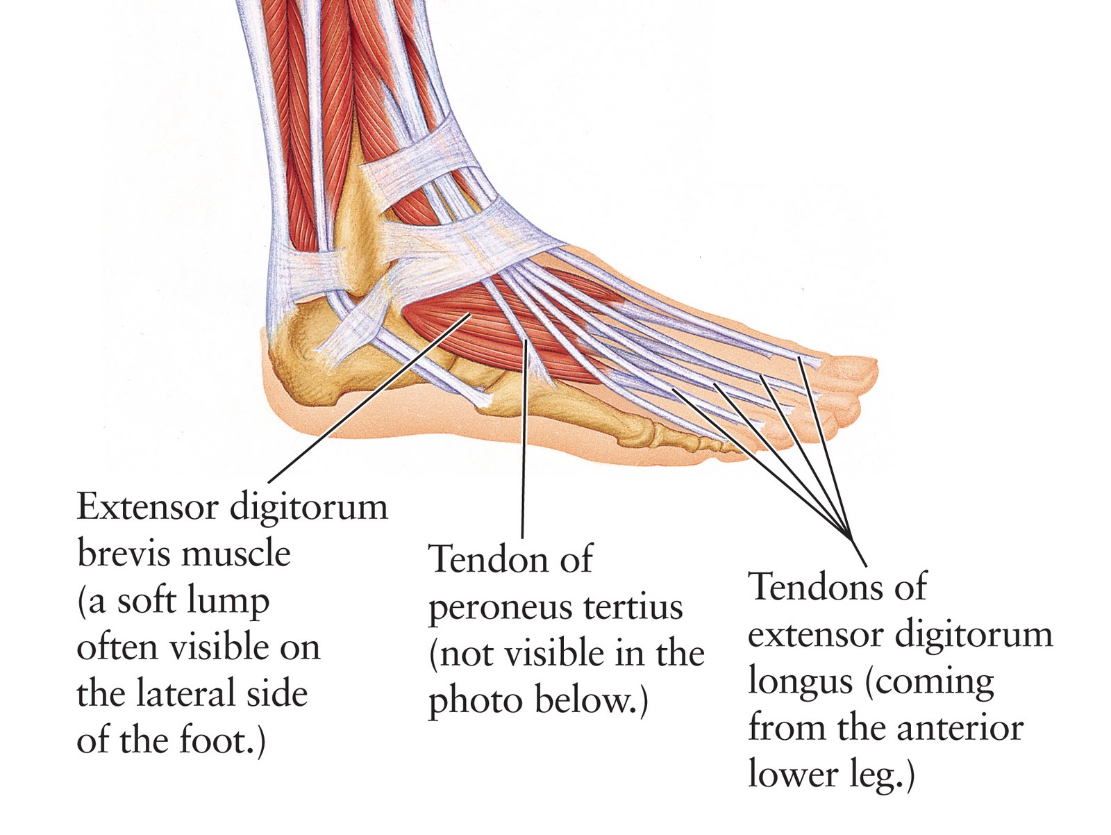
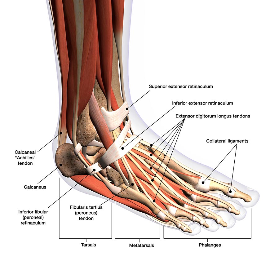
Human Anatomy for the Artist The Dorsal Foot How Do I Love Thee? Let Me Count Your Tendons
Tibia Fibula Talus Cuneiforms Cuboid Navicular Many of the muscles that affect larger foot movements are located in the lower leg. However, the foot itself is a web of muscles that can perform.

Foot pain looking up the chain
The Anatomy of Feet | Foot Diagrams, Physiology, and Pictures - Bilt Labs The Anatomy of Feet | Foot Diagrams, Physiology, and Pictures Made From The Molds Of Your Feet Active Designed for an active lifestyle. View Active Insoles Everyday Designed for normal day-to-day use. View Everyday Insoles Take Free Orthotics Quiz

Foot and Ankle Musculoskeletal Key
LABELED DIAGRAMS. Figure 1. Sections and Bones of the Foot A. Lateral (Left) B. Anterior (Right) Figure 2. Compartments of the Foot A. Cut Section through Mid-Foot. Figure 3. First Layer of the Foot A. Plantar View of Right Foot. Figure 4. Second Layer of the Foot A. Plantar View of Right Foot.
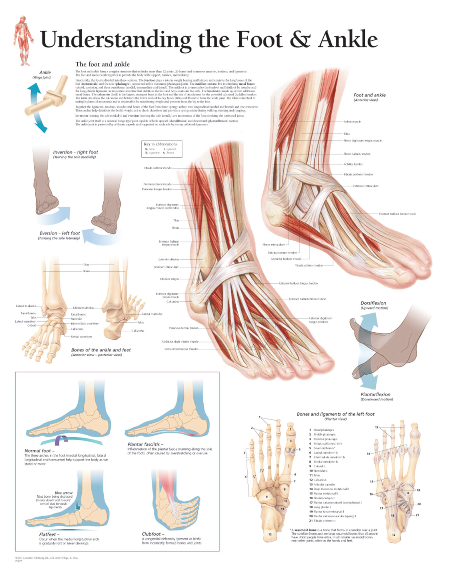
Understanding the Foot & Ankle Scientific Publishing
The 20-plus muscles in the foot help enable movement, while also giving the foot its shape. Like the fingers, the toes have flexor and extensor muscles that power their movement and play a large.

Anatomy of the Foot and Ankle OrthoPaedia
Common causes of foot pain include plantar fasciitis, bunions, flat feet, heel spurs, mallet toe, metatarsalgia, claw toe, and Morton's neuroma. If your feet hurt, there are effective ways to ease the pain. Some conditions specific to the foot can cause pain, less movement, or instability. Verywell / Alexandra Gordon.

Ankle and Foot Pain Massage Therapy Connections
When to see a doctor Summary The foot is an intricate part of the body, consisting of 26 bones, 33 joints, 107 ligaments, and 19 muscles. Scientists group the bones of the foot into the.

Chart of FOOT Dorsal view with parts name Vector image Stock Vector Image & Art Alamy
The phalanges create the toes. Each toe consists of three separate bones and two joints, except for the big toe, which has only two bones — distal and proximal phalanges — and one joint, like the.

Diagrams of Foot 101 Diagrams
Foot Diagram with Labels The foot is situated at the distal part of the lower limb. It is one structure that has undergone several evolutionary changes. The foot of humans has changed from grasping to a supporting structure. It supports the whole body weight while standing and also plays a vital role in locomotion.

Pin on Body (of) Work
Reading time: 20 minutes Recommended video: Ligaments of the foot [25:32] Comprehensive review of all major ligaments of the foot. Ankle joint Articulatio talocruralis 1/2 Synonyms: Talocrural joint The foot is the region of the body distal to the leg that is involved in weight bearing and locomotion.

Foot and Ankle Musculoskeletal Key
Figure 1: Bones of the Foot and Ankle Regions of the Foot The foot is traditionally divided into three regions: the hindfoot, the midfoot, and the forefoot (Figure 2). Additionally, the lower leg often refers to the area between the knee and the ankle and this area is critical to the functioning of the foot.